Diaa M. El-Mowafi - Zagagig University, Egypt
Ultrasonographic Versus Laparoscopic Control of Hysteroscopic Surgery
Diaa El-Mowafi
Associate Professor, Department of Obstetrics & Gynecology, Benha Faculty of Medicine, Egypt
Researcher & Educator, Wayne State Univerrsity, USA
Fellow, Geneva University, Switzerland
Abstract
This study was carried on 38 patients attending in Benha University Hospitals with different pathological intrauterine lesions that needed hysteroscopic surgery. The patients were divided into two groups A and B. Group A consists of 28 patients in whom transabdominal ultrasound was used as a control for the hysteroscopic surgery (17 endometrial resections, 5 myomectomies, 4 resections of uterine septum, and 2 divisions of intrauterine adhesions). Eight cases were done under cervical infiltration anaesthesia while the others were done under general anaesthesia. Group B consists of 10 patients in whom the traditional method of laparoscopic control under general anaesthesia was used during hysteroscopic surgery (6 endometrial resections, 2 myomectomies, 1 resection of uterine septum and 1 division of intrauterine adhesions). There were no complications in both groups a part from one perforation which needed no further management in group B due to obscuring of the field by adhesions from a previous pelvic operation. In one case of group A blanching of the peritoneal uterine coat occurred during endometrial resection but actual perforation did not occur. Ultrasound control of hysteroscopic surgery is recommended as it is easy, non-invasive, has no special complications or contraindications as for laparoscopy and allows the hysteroscopic surgery to be done under local cervical anaesthesia as an outpatient office procedure.
Introduction
The therapeutic indications of hysteroscopy extend to resection of submucous myomas and fibroid polyps, division of intrauterine septum and intrauterine synechae, endometrial resection and ablation, directed biopsy and removal of a lost intrauterine device (Lewis, 1989). Injection of polymers into the proximal fallopian tube for temporary or permanent sterilization may become a routine office practice. Transfer or retrieval of gametes and embryo may find an application (ACOG Technical Bulletin, 1994). Complications of hysteroscopy, particularly in surgical procedures, include uterine perforation, haemorrhage, gas embolism, fluid overload and infection (Rudigoz et al., 1994). Uterine perforation was reported early by Goldrath et al. (1981) in their first study of laser ablation and by Magos et al. (1989) in their series using the resectoscope in endometrial resection, while radiofrequency thermal ablation resulted in two cases of vesico-vaginal fistulae in the study done by Phipps et al. (1990). Mac Donald et al (1992) reported that perforation occurred during transcervical endometrial resection (TCRE). In 3 cases bladder was involved, in 2 ureteric damage occurred, and there were 2 major blood vessel injuries. They reported that 53% of perforations occurred in the first 5 cases performed by a surgeon. Six major complications occurred in the first 50 cases reported by Sturdee and Hoggart (1991) who stressed that the potential hazards of this operation must be appreciated and that training is essential . Laparoscopic control of intrauterine surgery was advised for many years (Siegler, 1983). In many women, when there is no suspected pelvic pathology, the laparoscope is considered as an invasive controlling tool carries additional risk and complications to the original hysteroscopic procedure (Shalev et al., 1994). Transabdominal ultrasound was used by other surgeons as an alternative aid to control hysteroscopic surgery (kohlenberg et al., 1994 and Querleu, 1994) .
Subjects and Methods
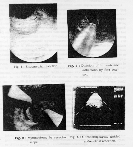
Thirty-eight patients attended the out patient clinic in Benha University Hospitals were admitted in this study. Twintv- three of them were complaining of abnormal uterine bleeding, 7 had 3 or more recurrent abortions, 5 were complaining of infertility one primary and 4 secondary for more than one year, 2 cases were presented with hypomenorrhoea, while one was presented with both secondary infertility and hvpomenorrhoea. The ages of the patients, were ranging between 21 and 45 years. Hysterosalpingography (HSG) was done for all patients except those presented with abnormal uterine bleeding for whom dilatation and curettage was done. HSG showed a picture suggestive of septate uterus which was documented later on by diagnostic laparoscopy in 5 cases and it showed filling defects in 10 cases. Endometrial carcinoma and adenomatous hyperplasia as well as cervical carcinoma were surely absent in the cases presented with abnormal uterine bleeding and included in the study as shown by their pathological reports. Our 38 patients were subjected to diagnostic then operative hysteroscoy in the same sitting with glycine 1.5% as a distension medium (fig. 1,2 and 3). The patients were divided into 2 groups (table 1) :
- Group A were subjected to operative hysteroscopy (Karl Storz, Germany) under transabdominal ultrasonography (Aloka-Sector Scanning, Japan) control (fig. 4). Eight cases of this group (2 septum, 2 intrauterine adhesions and 4 myomas) were done under cervical infiltration anaesthesia with 2% lignocaine. While the remaining 20 cases were done under general intubation anaesthesia.
- Group B were subjected to operative hysteroscopy under laparoscopic control (Karl Storz, Germany) with general intubation anaesthesia.
Results
In one case of laparoscopic controlled endometial resection by the resectoscope (karl Storz, Germany), a perforation in the posterior wall occurred due to obscuring of the field by adhesions from a previous pelvic operation (table II). It needed no further management. In one case of ultrasonographic controlled endometrial resection there was a suspicion of perforation. When lapasoscopy was performed immediately, only a whitish discoloration (blanching) of the uterine peritoneal coat was seen indicating no actual perforation but a heat transmitted from a very near end of the resectoscope. A difficulty to see a small posterior wall submucous myoma by ultrasound was encountered in one case. There were no complications in the other cases in both groups.

Discussion
A control of hysteroscopic surgery, mainly to guard against perforation of the uterus and its subsequent sequel, is essential particularly with the beginners (Siegler, 1983). Laparoscopic control is a virtual tool for this purpose particularly if another benefits could be achieved as evaluation of the tubal and peritoneal factors in a case of infertility or to exclude biconuate uterus in a hysterosalpingogram suggestive of septate uterus. But it is an invasive method, needs general anaesthesia, has its own complications as injury to the viscera or blood vessels and it is contraindicated in many situations as diaphragmatic hernia, large abdominal mass, ascitis, previous peritonitis and multiple pelvic surgery (Murphy, 1992). Disturbed anatomy or pelvic adhesions from a previous operation or infection may obscure the field as was in our complicated case.
Abdominal ultrasonographic control of hysteroscopic surgery is easy, non invasive, has neither contraindications nor operative complications and can have the advantage of guiding the hysteroscope towards the intrauterine lesion (kohlenberg et al., 1994). In the present study, the complication which was about to occur during ultrasonic control of endometrial resection (blanching) may be due to the thinness of the resectoscope terminal loop that it could not be seen easilv by the ultrasound. So a reasonable distance should be left between the visible end of the resectoscope and the internal margin of the uterine wall seen by ultrasound. These results are similar to that of Shalev et al. (1994) who reported that there were no complications, such as uterine perforation, during operative hysteroscopy controlled by ultrasound in 128 cases. The same was noticed in the study of kohlenberg et al. (1994) and Querieu (1994) who reported that the main advantages of transabdominal ultrasound as an aid to advanced hysteroscopic surgery are to direct the surgeon within the uterus to the site of pathology and to prevent inadvertent perforation of the uterine wall .
Table 1 : Hysteroscopic surgery done for the studied groups
| Hysteroscopic procedure | Tool used | Group A (n) | Group B (n) |
|
Endometrial resection |
resectoscope |
17 |
6 |
|
Myomectomy |
resectoscope |
5 |
2 |
|
Resection of septum |
resectoscope |
2 |
1 |
| Division of adhesions |
rigid scissor |
2 |
- |
|
fine scissor |
2 |
1 |
|
|
Total |
28 |
10 |
Group A Subjected to ultrasonographic controlled surgery
Group B Subjected to lapasoscopic controlled surgery
Table 2 : Complications in the two groups
|
Type of complication |
Group |
No. |
% |
|
Uterine perforation |
B |
1 |
10 |
|
Blanching of peritoneal coat |
A |
1 |
3.5 |
Conclusion
Abdominal ultrasonography control of hysteroscopic surgery is a safe, easy, non-invasive, and less operative time consuming than laparoscopic control. Hysteroscopic surgery with it can be an office procedure done under just an infiltrative cervical anaesthesia.
References
- ACOG technical bulletin (1994) Hysteroscopy. Int. J. Gynecol. Obstet. 45 (2) 175-180.
- Goldrath M., Tuller T. and Segal S. (1981) : Laser photovaporisation of endometrium for the treatment of menorrhagia. Am. J. obstet . Gynecol. 140 : 1419.
- Kohlenberg C., Pardy J. and Ellwood D. (1994) : Transabdominal ultrasound as an aid to advanced hysteroscopic surgery. Aust. NZ. J. Obstet. Gynecol. 34 (4) : 462 -464.
- Lewis B. (1989) : Hysteroscopy. In : Progress in Obstet. and Gynecol. Ed. Jobn Studd, Churchill Livingstone, London. vol. 7, pp 305-317.
- MacDonald R., Phipps J., and Singer A. (1992) Endornetrial ablation : a safe procedure. Gynecol. Endoscopy. I : 7-9.
- Magos A., Baumann R., Cheung k. and Turnbull A. (1989) : Transcervical resection of the endometrium in women with menorrhagia. Br. Med. J. 298:1209-1212 .
- Murphy A. (1992) : Diagnostic and operative laparoscopy. In: TeLinde's Operative Gvnecology. Ed Thompson J. and Rock J., J.B. Lippincott company, New York, p. 362 .
- Phipps J., Lewis B., Prior M. and Roberts T. (1990) : Experimental and clinical studies with radiofrequency induced thermal endometrial ablation for functional menorrhagia. Obstet. Gynecol. 76:876-881.
- Querleu D. (1994) : Echoguided resection of the septate uterus. J. Gynecol. Obstet . Biol. Reprod. 23 (5) 603 - 605.
- Rudigoz R., Marchal L., Gallien M. and Clement H. (1994): Complications of hysteroscopy. J. Gvnecol. Obstet. Biol. Reprod. 23 (5) : 503 - 510.
- Shalev E., Shimoni Y., and Peleg D. (1994) : Ultrasound controlled operative hvsteroscopy . J. Am. coll. surg. 179 (1): 70 - 71
- Sturdee D. and Hoggart B. (1991) : Problems with endometrial resection . Lancet 334 : 1474 - 1479.